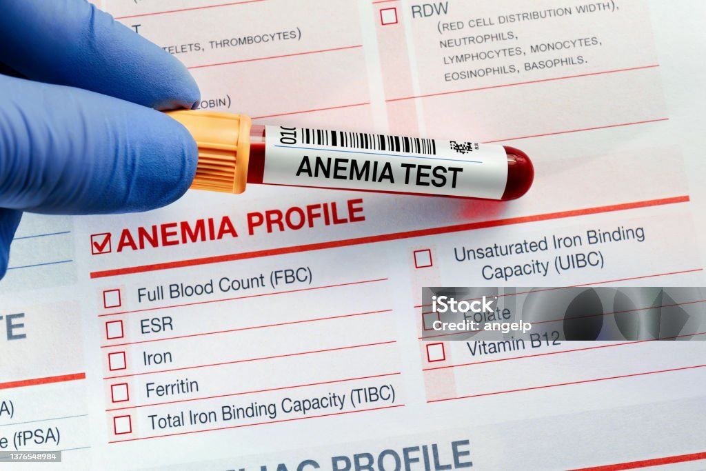
The Comprehensive Anemia Guide provides an authoritative exploration of anemia types, including insights into bone marrow functions and the intricacies of iron deficiency anemia.
From Pathophysiology to Clinical Management
Table of Contents
1. Definition and Overview
Anemia is a hematologic disorder characterized by:
- Hemoglobin (Hb) levels below 13 g/dL (men) or 12 g/dL (women)
- Reduced oxygen delivery to tissues due to:
- Decreased red blood cell (RBC) production
- Increased RBC destruction
- Blood loss
Key Pathophysiological Concepts
Erythropoiesis: Bone marrow process regulated by erythropoietin (EPO)
Hemoglobin synthesis: Requires iron, globin chains, and porphyrin ring
2. Epidemiology
Global Burden (WHO Data)
| Region | Prevalence | Affected Population |
|---|---|---|
| Worldwide | 24.8% | 1.62 billion people |
| Southeast Asia | 48.7% | Highest in women/children |
| Africa | 46.3% | Malaria-endemic regions |
Indian Scenario (NFHS-5)
| Population | Prevalence | Key Findings |
|---|---|---|
| Children (6-59m) | 67.1% | Iron deficiency predominant |
| Pregnant women | 52.2% | 30% of global maternal anemia |
| Elderly (>60y) | 42.1% | Chronic disease association |
3. Classification and Types
A. By RBC Morphology (MCV-Based)
Anemia Classification:
│
├── Microcytic (MCV<80fL)
│ ├── Iron Deficiency
│ ├── Thalassemia
│ ├── Anemia of Chronic Disease
│ └── Sideroblastic
│
├── Normocytic (MCV 80-100fL)
│ ├── Hemolytic
│ └── Acute Blood Loss
│
└── Macrocytic (MCV>100fL)
├── B12/Folate Deficiency
└── Myelodysplasia
│
├── Microcytic (MCV<80fL)
│ ├── Iron Deficiency
│ ├── Thalassemia
│ ├── Anemia of Chronic Disease
│ └── Sideroblastic
│
├── Normocytic (MCV 80-100fL)
│ ├── Hemolytic
│ └── Acute Blood Loss
│
└── Macrocytic (MCV>100fL)
├── B12/Folate Deficiency
└── Myelodysplasia
B. By Pathophysiological Mechanism
1. Hypoproliferative (Low reticulocytes)
- Nutritional deficiencies (Fe, B12, folate)
- Bone marrow failure
2. Hemolytic (High reticulocytes)
- Intrinsic RBC defects
- Extrinsic destruction
4. Iron Deficiency Anemia (IDA)
Pathogenesis
Stages of Depletion:
- Iron stores depletion (↓ ferritin)
- Iron-deficient erythropoiesis (↑ TIBC, ↓ serum Fe)
- Frank anemia (microcytic RBCs)
Diagnostic Approach
| Test | Result | Clinical Significance |
|---|---|---|
| Serum ferritin | <30 ng/mL | Earliest marker |
| Transferrin saturation | <16% | Functional iron deficiency |
| RBC morphology | Microcytic, hypochromic | Pencil cells, anisocytosis |
Management
Oral therapy: Ferrous sulfate 325 mg tid + vitamin C
IV options: Ferric carboxymaltose (15 mg/kg)
Monitoring: Reticulocyte crisis at 5-7 days, Hb ↑ by 1 g/dL/week
5. Anemia of Chronic Disease (ACD)
Key Features
Causes:
- Chronic infections (TB, HIV)
- Autoimmune disorders (RA, SLE)
- Malignancies
Pathophysiology
Inflammation → Cytokines → Hepcidin↑ → Iron Sequestration
Cytokines → EPO↓ → Marrow Suppression
Cytokines → EPO↓ → Marrow Suppression
Diagnostic Criteria
| Parameter | ACD Pattern | IDA Pattern |
|---|---|---|
| Serum iron | ↓ | ↓↓ |
| Ferritin | Normal/↑ | ↓ |
| TIBC | ↓ | ↑ |
Treatment
- Underlying disease control
- ESA therapy (Epoetin alfa 50-100 U/kg TIW)
- IV iron if functional deficiency present
6. Thalassemia Syndromes
Genetic Basis
| Type | Defect | Clinical Severity |
|---|---|---|
| α-thalassemia | α-globin gene deletion | Silent → HbH disease |
| β-thalassemia | β-globin mutations | Minor → Major |
Diagnostic Workup
- CBC: Marked microcytosis (MCV often <70fL)
- Hb electrophoresis:
- β-thalassemia: ↑HbA2 (>3.5%), ↑HbF
- α-thalassemia: HbH inclusions
- Genetic testing: Definitive diagnosis
Management
Transfusion-dependent:
- Regular PRBC + iron chelation
- Target Hb >9.5 g/dL
Curative: Allogeneic HSCT
7. Sideroblastic Anemia
Classification
- Hereditary: X-linked (ALAS2 mutations)
- Acquired:
- Myelodysplastic syndrome (MDS-RS)
- Toxins (lead, alcohol)
Pathognomonic Findings
Bone marrow:
- Ring sideroblasts (>15% of erythroblasts)
- Prussian blue stain shows perinuclear iron
Blood:
- Dimorphic RBC population
- Basophilic stippling
Treatment
- Pyridoxine: 100-200 mg/day (X-linked forms)
- Chelation therapy for iron overload
- HSCT for MDS-associated cases

8. Diagnostic Algorithms
Microcytic Anemia Workup
[MCV<80]
│
├── Ferritin
│ ├── Low → Iron Deficiency
│ └── Normal/High → Hb electrophoresis
│ ├── Abnormal → Thalassemia
│ └── Normal → ACD or sideroblastic
│
├── Ferritin
│ ├── Low → Iron Deficiency
│ └── Normal/High → Hb electrophoresis
│ ├── Abnormal → Thalassemia
│ └── Normal → ACD or sideroblastic
9. Therapeutic Guidelines
Blood Transfusion Thresholds
| Clinical Scenario | Hb Threshold |
|---|---|
| Stable outpatient | <7 g/dL |
| Cardiovascular disease | <8 g/dL |
| Active bleeding | <9 g/dL |
10. Complications
Organ-Specific Effects
| System | Complication | Management |
|---|---|---|
| Cardiac | High-output failure | Slow transfusions + diuretics |
| Endocrine | Hypogonadism (thalassemia) | Hormone replacement |
| Hepatic | Cirrhosis (iron overload) | Phlebotomy/chelation |
11. Prevention Strategies
Population-Level
- Iron fortification: Wheat flour, salt
- Malaria prophylaxis: In endemic regions
- Genetic counseling: For thalassemia carriers
High-Risk Groups
| Population | Intervention |
|---|---|
| Pregnant women | 60 mg elemental Fe + 400 μg folate |
| Elderly | Annual CBC + nutritional screen |
Key Takeaways
- Anemia is classified by RBC size (MCV) and underlying cause
- Iron studies differentiate between IDA, ACD, and sideroblastic anemia
- Thalassemia requires Hb electrophoresis for diagnosis
- Treatment must address both symptoms and underlying etiology